Mostrar el registro sencillo del ítem
dc.contributor.author
Costantino, Valeria Victoria

dc.contributor.author
Bocanegra, María Victoria

dc.contributor.author
Cacciamani, Valeria

dc.contributor.author
Gil Lorenzo, Andrea Fernanda

dc.contributor.author
Benardon, María Eugenia

dc.contributor.author
Vallés, Patricia G.
dc.date.available
2020-07-22T20:15:33Z
dc.date.issued
2019-10
dc.identifier.citation
Costantino, Valeria Victoria; Bocanegra, María Victoria; Cacciamani, Valeria; Gil Lorenzo, Andrea Fernanda; Benardon, María Eugenia; et al.; Losartan through Hsp70 Avoids Angiotensin II Induced Mesenchymal Epithelial Transition Proximal Tubule Cells from Spontaneously Hypertensive Rats; Karger; Cellular Physiology and Biochemistry; 53; 4; 10-2019; 713-730
dc.identifier.issn
1015-8987
dc.identifier.uri
http://hdl.handle.net/11336/109972
dc.description.abstract
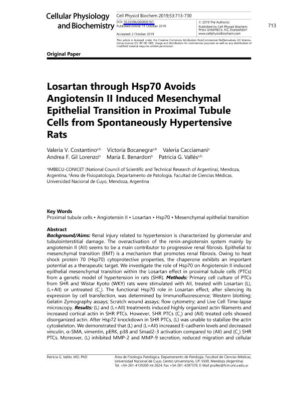
Renal injury related to hypertension is characterized by glomerular and tubulointerstitial damage. The overactivation of the renin-angiotensin system mainly by angiotensin II (AII) seems to be a main contributor to progressive renal fibrosis. Epithelial to mesenchymal transition (EMT) is a mechanism that promotes renal fibrosis. Owing to heat shock protein 70 (Hsp70) cytoprotective properties, the chaperone exhibits an important potential as a therapeutic target. We investigate the role of Hsp70 on Angiotensin II induced epithelial mesenchymal transition within the Losartan effect in proximal tubule cells (PTCs) from a genetic model of hypertension in rats (SHR). Methods: Primary cell culture of PTCs from SHR and Wistar Kyoto (WKY) rats were stimulated with AII, treated with Losartan (L), (L+AII) or untreated (Cc ). The functional Hsp70 role in Losartan effect, after silencing its expression by cell transfection, was determined by Immunofluorescence; Western blotting; Gelatin Zymography assays; Scratch wound assays; flow cytometry; and Live Cell Time-lapse microscopy. Results: (L) and (L+AII) treatments induced highly organized actin filaments and increased cortical actin in SHR PTCs. However, SHR PTCs (Cc ) and (AII) treated cells showed disorganized actin. After Hsp72 knockdown in SHR PTCs, (L) was unable to stabilize the actin cytoskeleton. We demonstrated that (L) and (L+AII) increased E-cadherin levels and decreased vinculin, α-SMA, vimentin, pERK, p38 and Smad2-3 activation compared to (AII) and (Cc ) SHR PTCs. Moreover, (L) inhibited MMP-2 and MMP-9 secretion, reduced migration and cellular displacement, stabilizing intercellular junctions. Notably, (L) treatment in shHsp72 knockdown SHR PTCs showed results similar to SHR PTCs (Cc ). Conclusion: Our results demonstrate that Losartan through Hsp70 inhibits the EMT induced by AII in proximal tubule cells derived from SHR.
dc.format
application/pdf
dc.language.iso
eng
dc.publisher
Karger

dc.rights
info:eu-repo/semantics/openAccess
dc.rights.uri
https://creativecommons.org/licenses/by-nc-sa/2.5/ar/
dc.subject
Angiotensin II
dc.subject
Hsp70
dc.subject
Losartan
dc.subject
Mesenchymal epithelial transition
dc.subject
Proximal tubule cells
dc.subject
Hsp70
dc.subject
Losartan
dc.subject
Mesenchymal epithelial transition
dc.subject.classification
Bioquímica y Biología Molecular

dc.subject.classification
Ciencias Biológicas

dc.subject.classification
CIENCIAS NATURALES Y EXACTAS

dc.title
Losartan through Hsp70 Avoids Angiotensin II Induced Mesenchymal Epithelial Transition Proximal Tubule Cells from Spontaneously Hypertensive Rats
dc.type
info:eu-repo/semantics/article
dc.type
info:ar-repo/semantics/artículo
dc.type
info:eu-repo/semantics/publishedVersion
dc.date.updated
2020-06-30T14:23:28Z
dc.journal.volume
53
dc.journal.number
4
dc.journal.pagination
713-730
dc.journal.pais
Suiza

dc.journal.ciudad
Basel
dc.description.fil
Fil: Costantino, Valeria Victoria. Universidad Nacional de Cuyo; Argentina. Consejo Nacional de Investigaciones Científicas y Técnicas. Centro Científico Tecnológico Conicet - Mendoza. Instituto de Medicina y Biología Experimental de Cuyo; Argentina
dc.description.fil
Fil: Bocanegra, María Victoria. Universidad Nacional de Cuyo; Argentina. Consejo Nacional de Investigaciones Científicas y Técnicas. Centro Científico Tecnológico Conicet - Mendoza. Instituto de Medicina y Biología Experimental de Cuyo; Argentina
dc.description.fil
Fil: Cacciamani, Valeria. Consejo Nacional de Investigaciones Científicas y Técnicas. Centro Científico Tecnológico Conicet - Mendoza. Instituto de Medicina y Biología Experimental de Cuyo; Argentina
dc.description.fil
Fil: Gil Lorenzo, Andrea Fernanda. Universidad Nacional de Cuyo; Argentina
dc.description.fil
Fil: Benardon, María Eugenia. Universidad Nacional de Cuyo; Argentina
dc.description.fil
Fil: Vallés, Patricia G.. Consejo Nacional de Investigaciones Científicas y Técnicas. Centro Científico Tecnológico Conicet - Mendoza. Instituto de Medicina y Biología Experimental de Cuyo; Argentina. Universidad Nacional de Cuyo; Argentina
dc.journal.title
Cellular Physiology and Biochemistry

dc.relation.alternativeid
info:eu-repo/semantics/altIdentifier/doi/http://dx.doi.org/10.33594/000000167
dc.relation.alternativeid
info:eu-repo/semantics/altIdentifier/url/https://www.cellphysiolbiochem.com/Articles/000167/
Archivos asociados
